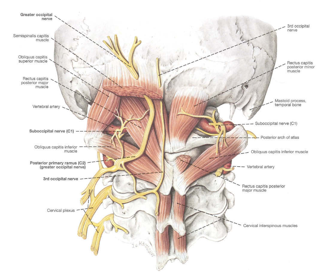
Occipital Neuralgia (Occipital Neuralgia ) Is a condition of severe pain in the head that extends from the back of the head to the neck. The distribution of pain follows the innervation of the greater occipital and third occipital, namely the sensory nerves that run from the upper neck to the back of the head. The pain can be throbbing, sharp, stabbing.
Pain that arises due to irritation and inflammation of the occipital nerves caused by various conditions such as trauma to the neck and back of the head, excessive muscle spasms, arthritis in the cervical spine and structural changes in the upper cervical spine. Usually also accompanied by the presence of other diseases such as Diabetes Mellitus, infection, inflammation and blood disorders.

*Location of the Greater Occipital Nerve and Third Occipital Nerves
Although the complaints found in patients vary widely, almost all locations of pain are along the upper neck, the junction of the neck and the bones of the head to the back of the skull. The pain can be on one side or on both sides of the head and upper neck bones. Pain can sometimes radiate to the front of the head.
However, anamnesis history of pain in patients accompanied by an accurate physical examination can determine the diagnosis of this disease correctly. Physical examination is carried out by pressing on the distribution of the occipital nerve course. If there is severe pain, the diagnosis can be made. Usually also accompanied by spasms in the neck muscles. Supporting examinations such as X Ray, CT Scan and MRI are needed if there is a suspicion of structural abnormalities of the cervical spine and the back of the head.
Medical treatment for this disease varies greatly. Starting with painkillers and muscle relaxant therapy, physiotherapy with massage and heating, rest, injections of local anesthetics and steroids. Usually, patients take painkillers for a long time and then consult a pain specialist.
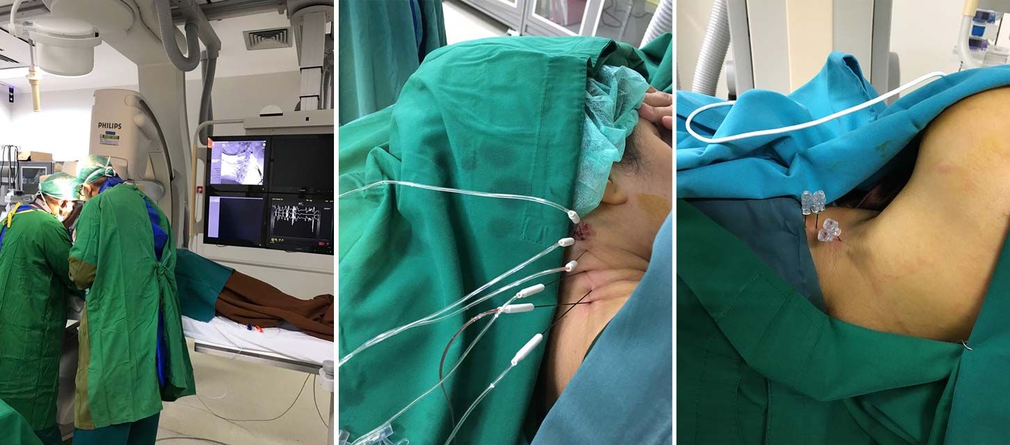
If the response to the above treatment variations fails and the pain returns very repeatedly, then therapy is carried out in the form of Rhizotomy, which blunts the course of the nerve roots so that it can reduce pain immediately, then proceed with the process of neurolysis, namely by providing heat stimulation and other chemical reactions so that it inhibits the transmission of pain directly on the course. the nerves. This minimally invasive action is carried out using a needle and then connected to a radiofrequency machine and carried out in the Cathlab room which is used to get optimal visualization of the course of the procedure in an updated manner so that it is safer and guides the needle towards proper and accurate nerve distribution.

*Example of Neurolysis Ablation with Radiofrequency
The article was written by a Neurosurgeon Specialist at EMC Sentul Hospital.
